Echinococcosis is a well‐known parasitic disease of
tapeworms of the echinococcus type.
the disease may occur in most areas of the world but is
particularly prevalent in africa, south america and
asia, where it may affect 5‐10 % of the population. the
economic cost of the disease is huge and is estimated to
be around 3 billion usd a year (1).
although the disease is relatively rare in western europe,
due to immigration of populations from countries where
the disease is endemic and increased travelling, this
tropical disease may resurge in western countries.
most physicians are aware of the abdominal,
pulmonary and probably also brain manifestations of
the disease. bone and soft tissue manifestations,
however, are often neglected in the differential
diagnosis in nonendemic countries.
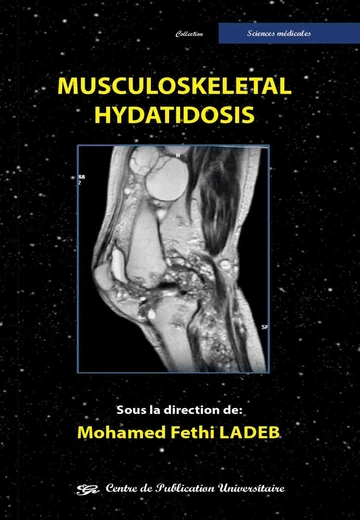
in this regard, this updated monograph addressing the
imaging characteristics of musculoskeletal
echinococcosis is a much‐wanted survey filling the
gaps of our knowledge on the disease.
the book is written by a team of experts in the field lead
by professor mohamed fethi ladeb and his collaborators. after a comprehensive overview of the pathogenesis,
histopathology and clinical manifestations of
echinococcosis, all details on imaging of musculoskeletal
manifestations of the disease are extensively discussed.
the book is richly illustrated by well‐selected highquality
images and covers all imaging techniques.
the book reads very fluently and is a “must‐have” on
the shelf of every library.
we are convinced that this work will be a very useful
tool not only for musculoskeletal radiologists but also
for all other colleagues involved in the diagnosis and
management of musculoskeletal disorders as well as
for clinicians of internal medicine particularly those
practicing infectious and tropical diseases.
we cordially congratulate the editors, professor
mohamed fethi ladeb and his team, for this excellent job,
and it is our great privilege to recommend this book as a
reference work on musculoskeletal echinococcosis.

